Central Vein Occlusion is the second most common vascular disease after diabetic retinopathy. Vascular disease refers to disorders affecting blood vessels, crucial in transporting oxygen and nutrients throughout your body while eliminating waste from your tissues. To understand CRVO treatment, classify it and know the three distinct units per the occlusion’s anatomic level.
Central retinal vein occlusion ( CRVO)– involves the occlusion at the level of or posterior to the lamina cribrosa.
Hemiretinal vein occlusion( HRVO) is at the disc involving inferior or superior hemiretina.
Branch retinal vein occlusion ( BRVO)– involves occlusion of tributary vein mainly at the site of arteriovenous crossing.
What happens in Retinal Vein Occlusion?
Retinal Vein Occlusion is a vascular disease wherein the veins that drain the blood from the retina get partially or entirely blocked. The retina is the white layer at the back of the eyeball that helps light entering the eye get translated into images. The blockage prevents the draining of blood from leaving the retina. This complication occurs due to increased blood pressure and swelling in your eyes. Prompt Central retinal vein occlusion treatment can prevent and minimize vision loss.
What is the significant risk factor associated with CRVO ?
The primary risk factor associated with CRVO includes open-angle glaucoma. Individuals with CRVO in one of the eyes are at a higher risk of developing CRVO in a fellow eye. Systemic disorders for CRVO include increasing age, diabetes mellitus and hypertension.
Read More: All About CRVO treatment And Ayurveda Remedies
What are the symptoms of CRVO?
In the case of Mild CRVO, many instances may show no symptoms. However, many patients with CRVO report symptoms such as blurry or irregular vision due to swelling of the center part of the retina, also known as the macula.
Patients with severe CRVO symptoms show secondary complications such as glaucoma, a disease which is characterized by increased pressure in the eye. They also experience pain, redness, irritation and other types of problems.
What are the different causes of retinal vein occlusion?
RVO is caused by disruption to normal blood flow through the retinal vein causing this condition. The disruption may happen due to:
- A blood clot
- A deceleration of blood flow.
- Contraction of the retinal vein at the point where it intersects paths with the retinal artery.
The retinal artery provides oxygen-rich blood to the retina. However, the retinal artery may grow stiff due to aging or plaque formation. It may press on the retinal vein and cause harm to the inner lining of your retinal vein, leading to circumstances that increase the likelihood of blood clot formation.
What do Stats reveal about CRVO?
As we know, Retinal vein occlusion is the second most common disorder impacting the retina after diabetic retinopathy. Still, researchers estimate that Retinal vein occlusion ( RVO) impacts more than 16 million people globally. If we talk about the types of RVO, Central retinal vein occlusion is believed to affect 1 to 4 individuals in 1,000 people, whereas Branch retinal vein occlusion impacts 6 to 12 individuals in 1,000 people.
What diagnosis can provide helpful information about CRVO?
Optical Coherence Tomography: OCT helps to confirm and measure the extent of severity of macular edema, evaluate the photoreceptor layer and monitor the response to CRVO treatment. The treatment of OCT measurement often becomes the guide treatment. Patients’ age directs the systemic diagnosis of patients with CRVO and co-existing risk factors.
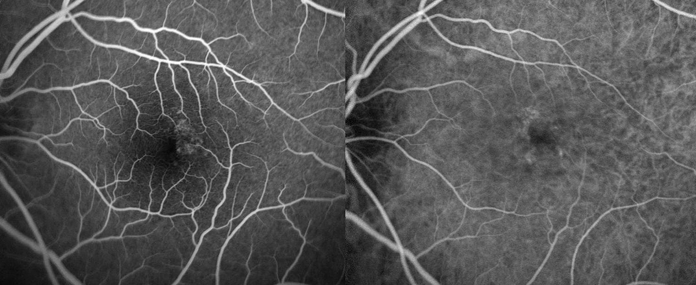
Fluorescein angiography ( FA) – The FA allows evaluation of the capillary extent of capillary nonperfusion and the degree of macular ischemia.
Complications Associated with Retinal vein occlusion
Retinal vein occlusion may lead to various complications such as:
- Cystoid macular edema: In this complication, there is swelling in the retina’s (macula) center. It can lead to blurry vision or loss of vision.
- Neovascularization of the eye: Abnormal blood vessels are formed in different parts of the eyes, mainly the iris (rubeosis iridis). This occurs in approximately 1 in 4 people with RVO. Abnormal blood vessels less naturally form in the retina.
- Bleeding in the eye (vitreous haemorrhage) occurs when blood leaks into the vitreous humour, a gel-like material that fills the eyeball. It occurs from the construction of abnormal blood vessels prone to leaking.